
Building on its history of excellence in medical physics and accelerator technology, the University of Saskatchewan is well-positioned to lead the country in nuclear medicine, playing a lead role in biomedical imaging and nuclear research, development and training.
These initiatives—ranging from bio-medical imaging at the Canadian Light Source to plant, animal and human imaging research and development at the Sylvia Fedoruk Canadian Centre for Nuclear Innovation—constitute a growing USask bio-imaging cluster which enhances and supports an array of expertise and collaborative opportunities in plant, animal and human health research through the College of Medicine, Western College of Veterinary Medicine, College of Agriculture and Bioresources, Saskatoon Cancer Centre, Royal University Hospital, the Vaccine and Infectious Disease Organization-International Vaccine Centre (VIDO-InterVac), the Global Institute for Food Security, and the Global Institute for Water Security.
Sylvia Fedoruk Canadian Centre for Nuclear Innovation (Fedoruk Centre)
Advancing R&D in Nuclear Medicine, Materials, Energy and Environment
Sylvia Fedoruk Canadian Centre for Nuclear Innovation (Fedoruk Centre) is a non-profit subsidiary and research centre of the University of Saskatchewan
Through partnership investments in nuclear science and technology that support USask's research and academic mission, the Fedoruk Centre places Saskatchewan among global leaders in nuclear research, development and training.
The Fedoruk Centre builds on USask's historical strengths in areas such as nuclear medicine, accelerator technology and materials sciences research by funding academic programming and research and development projects in three target impact areas:
- Nuclear imaging tools and methods to advance life sciences, agri-biotechnologies and medicine;
- Material sciences, through nuclear techniques, to improve energy, health, and transportation; and
- Understanding the practical and social aspects of nuclear energy, to inform decision-making towards a clean, sustainable future
For more information, visit: Sylvia Fedoruk Canadian Centre for Nuclear Innovation
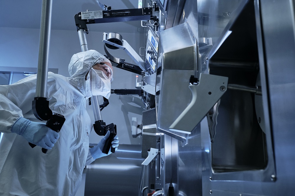
Saskatchewan Centre for Cyclotron Sciences
Accelerating Innovation for Medical Diagnosis and Treatment
This state-of-the-art particle accelerator of the University of Saskatchewan, managed by the Fedoruk Centre, supports innovation in nuclear imaging and therapy in living specimens: plants, animals and humans.
This unique-in-Canada facility featuring a high-energy cyclotron, radiopharmaceutical production facility, and radiochemistry laboratories enables research in isotope use and detection technologies for medical diagnosis and treatment, and training operators of accelerators and future researchers in the area of accelerator physics.
The SCCS is currently being used to study a diverse range of subjects including developing the next generation of molecular imaging agents for cancer diagnostics, developing new targeted smart drugs to fight cancer and antibiotic-resistant infections, and imaging microbial and root activity in soil ecosystems.
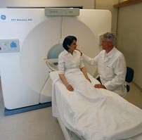
The cyclotron is a class II nuclear facility certified by Health Canada for production of radiopharmaceuticals, and produces short-lived radioactive isotopes for use in Saskatchewan’s first PET-CT (Position Emission Tomography – Computed Tomography) scanner. Since 2016, SCCS has provided medical isotopes for nuclear imaging scans of more than 5,000 Saskatchewan patients at Royal University Hospital, as well as for diagnosing and treating cancer in Alberta and Manitoba.
By 2021, the SCCS expects to bring 10 new products, services or technologies to the marketplace, generate at least 60 patents or publications, and train at least 50 new highly qualified people.
For more information, visit: Saskatchewan Centre for Cyclotron Sciences
Bio-Medical Imaging and Therapy (BMIT) Beamlines at the Canadian Light Source
Sharper Image Brings Tiny Tumors to Light
University of Saskatchewan (USask) scientists are using synchrotron light to develop technology that provides unprecedented image quality and will lead to better diagnosis and treatment of diseases such as cancer, cystic fibrosis, and osteoporosis.
Their state-of-the-art tool is the BioMedical Imaging and Therapy (BMIT) beamline facility at the USask Canadian Light Source synchrotron. BMIT is unique in North America and one of only a handful of such facilities in the world. It currently undertakes about 60 unique experiments per year with scientists from across Canada and around the globe.
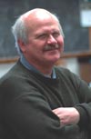
Initially established by USask medical imaging researcher Dean Chapman and fully operational since 2015, BMIT is capable of imaging and therapy research benefitting human and animal health.
The remarkable advantage of synchrotron X-ray imaging is the ability to detect structural changes that can’t be seen with other techniques such as conventional X-ray machines or MRI. This can be used to answer important questions about where diseases originate and follow their progression.
One technique invented by Chapman and used at BMIT - diffraction-enhanced imaging (DEI) - uses synchrotron X-rays to capture images of soft tissues that are invisible to normal X-ray machines.
With DEI, scientists can detect and measure how synchrotron-generated X-rays refract and scatter when they pass through various tissues, creating high-resolution images of muscles and organs that clearly reveal details such as the extent of cancer tumors in breast tissue or damaged areas in lungs. DEI can provide 33 times greater image contrast and dramatically less radiation exposure than regular X-rays. The technique promises better cancer detection, particularly in mammography.
Phase contrast imaging is another technique used regularly at BMIT. The technique which is also sensitive to soft tissues and detects X-ray absorption and refraction by tissue, can be used to detect structural changes down to the size of the cells (1 micron).
Since becoming operational, BMIT has been used by Canadian scientists in hundreds of research projects including in physiology, human and veterinary medicine, and many other areas.
Data collected at BMIT beamlines have now contributed to over 300 publications, including journal articles, graduate students' theses, conference proceedings and book chapters.
Recent health research highlights from BMIT:
- Developing microbeam radiation therapy for inoperable cancer
- Helping people hear by imaging the complex internal anatomy of the ear
- Improving treatment for cystic fibrosis
The X-ray techniques available at BMIT are also frequently used by a diverse group of researchers in other fields such as pharmaceutics, agriculture, advanced materials, renewable energy, rechargeable batteries, paleontology, and oil and gas.
Canadian Isotope Innovations Corp.
Advancing the Search for a Safe and Reliable Supply of Medical Isotopes
Producing medical isotopes cheaply and reliably, without using a nuclear reactor or highly enriched uranium, is the aim of Canadian Isotope Innovations Corp (CIIC), a company commercializing technology initially developed at the Canadian Light Source (CLS) synchrotron, a national research facility of the University of Saskatchewan.
The first facility of its kind in the world, CIIC uses a linear accelerator facility within the CLS, which was funded by the Canadian federal and Saskatchewan provincial governments. CIIC also uses the university’s cyclotron, managed by the Fedoruk Centre at University of Saskatchewan, for processing.
Unlike traditional medical isotope production methods, CIIS uses a linear accelerator (linac) that does not produce radioactive waste and does not use enriched uranium.
A high energy electron beam from a linac hits a dense target, producing high energy X-rays. These X-rays then strike atoms of a second target, transforming some of the target atoms to the desired isotope. After bombarding the targets for a few hours to days, the irradiated targets are then processed to extract the desired isotope in a form that CIIC distributes to radiopharmacies and nuclear medicine clinics. The isotopes are attached to drugs to form radiopharmaceuticals for specific diagnostic tests or cancer therapies.
Two products are ideally suited to electron linac production:
- Technetium-99 (Tc-99m), required by nuclear medicine physicians for early stage diagnosis of disease (such as cancer & cardiovascular disease), is the most common radioisotope used in medical diagnosis procedures. In combination with imaging devices, the gamma rays emitted by the isotopes can be used to study the dynamic processes taking place in various parts of the body. Nearly 17 million procedures using Tc-99m are performed each year in the United States and 1.3 million are performed in Canada.
- Copper 67 (Cu 67) is an emerging radioisotope used for radionuclide therapy (RNT) or radiotherapy. Some cancerous growths can be controlled or eliminated by irradiating the area containing the growth. The radioisotope that generates the radiation can be localized in an affected organ in the same way as for diagnosis – through a radioisotope following its usual biological path, or through the isotope being attached to a suitable biological compound.
CIIC continues to develop the processes to enable commercial distribution to Canadian and international radiopharmacies for both radioisotopes.
If the approach proves technically and economically feasible, accelerator clusters in one or more locations in Canada would produce molybdenum-99 (Mo-99), which would be transported to hospitals for recovery of the Tc-99m using a radionuclide separator.
For more information, visit: http://isotopeinnovations.com/